|
|
|
|
|
|
|
|
|
|
|
|
|
|
|
|
|
|
|
|
|
|
|
|
|
|
|
|
|
|
|
|
|
|
|
|
|
|
|
|
|
|
|
|
|
|
|
|
|
|
|
|
|
|
|
|
Towards an Integrated Approach of sEMG Utilization: Quantative Protocols of Assessment and Biofeedback
Gabriel E. Sella,
M.D., M.P.H., M.Sc.
Introduction Surface electromyography is a computerized electrophysiological technology that can be utilized in an array of approaches. It is an objective tool for assessment and diagnosis of muscular electrical activity in health and disease. It is an objective electronic monitor during the course of physical and occupational therapy for most neuromuscular conditions. sEMG is similar to a voltmeter device. It measures the summation of passage of multiple units of action potentials (m.u.a.p) between two points on a muscle belly from the vantage point of the skin surface. It can also measure the frequency of passage of the m.u.a.p.s. The summation of the action potential passage shows up as an amplitude form, i.e. the greater the number of passing action potentials, the higher the amplitude of the micro-voltage. The classic distance between the two surface electrodes is two centimeters. If the electrodes are placed at a larger distance, the number of action potentials coursing between the two points will be larger and thus the amplitude will be higher. sEMG Can Measure Parameters Of Muscular Activity in Health and Disease The electrical muscular activity in a normal muscle will include three parameters:
The electrical activity is normally within 10% of amplitude or "area beneath the curve" between two normal contra-lateral muscles, which are activated through the same range of motion and against the same resistance. The activity can be tested through a number of repetitions, in order to determine a number of phenomena. These are: Passage from initial activity of fast twitch muscle fibers to slow twitch fibers (spectral analysis) over time, substitution patterns (especially after 10 seconds of sustained contractions) and fatigue phenomena, sometimes observable with the median frequency parameter (Hz).
The basic principles of the "Sella sEMG Protocols" are as follows:
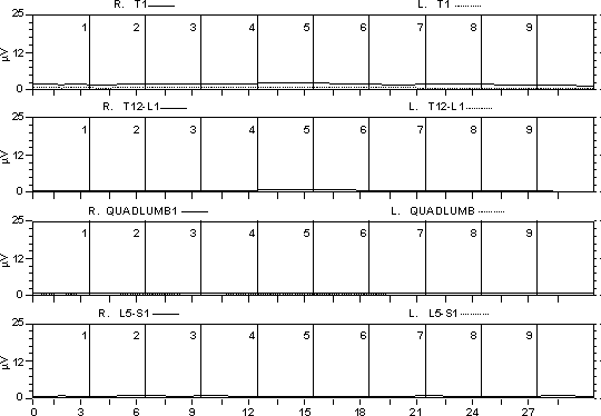
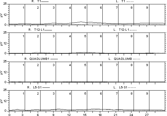
At least 5 repetitions of motion through the R.O.M. are needed in order to establish the repeatability and statistical validity of the compliance of the person tested for the requirements of the testing. Testing of the baseline activity, at rest, needs to be performed initially, i.e. before the R.O.M. protocol, between periods of muscular activity through the R.O.M. and at the end of testing. Testing can be done in three different modalities with regards to gravity and resistance. The first modality is that of the muscle motion through the R.O.M. without any resistance or conscious effort. The second modality is that of the muscle motion through the R.O.M. while the person exerts maximal voluntary contraction. The third modality involves a set resistance against the muscle's motion through the R.O.M. (note, a variant of this modality involves an isometric contraction, i.e. with a set resistance so large that the muscle contracts without being able to move the joint in space). Statistical analysis needs to be done. The computer software should be able to produce statistical data which enables the investigator or clinician to interpret the sEMG results. This data should include: Consistency of baseline values before, during and after testing the range of motion. Such baseline values should normally be Initial Rest:
Figure 3 Final Rest:
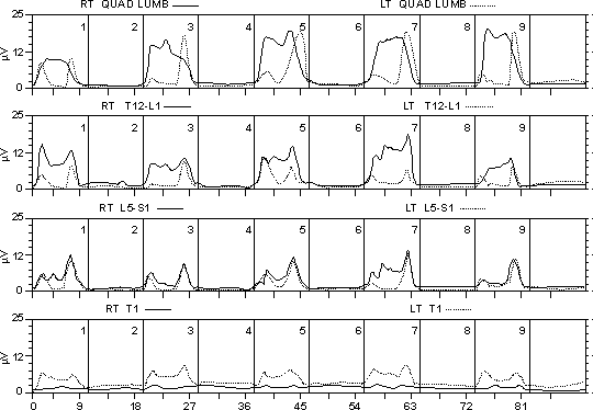
The same resting tonus is obtained during resting periods between back rotations to the right: Resting Periods:
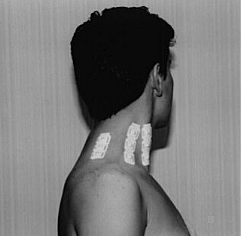
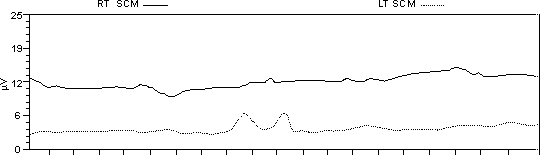
Figure 5 The following graph illustrates the fact that normal resting values may co-exist with abnormal ones, such as with muscle spasm. The example, which pertains to whiplash accident, shows the right SCM as having a resting value of approximately 12mV, while the left SCM has a resting value of approximately 4mV:
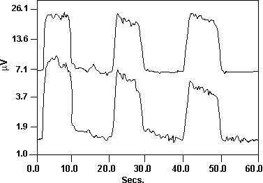
Figure 6 Consistency of motion values during the R.O.M. of the muscles tested such that the values should be normally >4mV in amplitude and within 10% for each contra-lateral pair of muscles through any number of repetitions of the R.O.M. The same should apply for the areas beneath the curve of motion through any R.O.M. The graph below illustrates this point on the right and left SCM during neck flexion. The maximal amplitudes during activity are above 4mV and are very close to one another for the contra-lateral muscles.
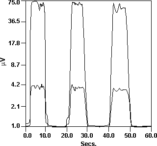
Figure 7 Hypertonus is an abnormal pattern of muscular behavior defined as a harmonic motion where the baseline or rest value is repeated >4mV through the R.O.M.. By definition, the difference in amplitude of activity during rest and voluntary muscular contraction should be >10% for any given muscle repetition through the R.O.M. Abnormal patterns, such as hypertonus, should follow the same rules of statistical analysis as described in principle 6. The graph below illustrates this point on the extensor carpi radialis during wrist extension:
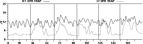
Figure 8 The maximal amplitudes during rest are above 4mV on the right side. The left resting period is <4mV. There is a clear tendency to return to rest for the muscles involved. However, this is not the case in the muscles affected by the injury. Other muscles, tested at the same time, show normal resting values. Muscle spasm is an abnormal pattern of muscular activity through the R.O.M. It is defined as a level of activity >4mV in which there is no discernible difference in the amplitude of contraction during rest or motion. The statistical data should demonstrate that the difference between the amplitude of activity, at rest and during motion, is <10% for any given muscle in spasm through any number of contractions through the R.O.M. The graph below illustrates the point on the right upper trapezius during neck flexion. The maximal amplitudes during rest are above 4mV for the right upper trapezius in spasm. There is no tendency to return to rest for the muscles involved, however, this is not the case in the muscles affected by injury. The left upper trapezius, tested at the same time, shows normal resting values.
Figure 9 Muscular contracture, or silence, is an abnormal pattern of muscular activity through the R.O.M. It is defined as a level of activity close to 1mV in which there is no discernible difference in the amplitude of contraction during rest or motion. The statistical data should demonstrate that the difference between amplitude of activity at rest and during motion is <10% for any given muscle in spasm through any number of contractions through the R.O.M. Muscular contracture should be ruled out from being a technical artifact. Statistical parameters of interpretation need to include at least the following:
The median frequency (Hz) difference between two normal contra-lateral muscles can be considered to be normal if the difference is <10% for any given contraction through the R.O.M. All the considerations described above apply for this parameter. It is expected, clinically and statistically, that there should be no significant differences for the parameters described above in normal muscles. Symptom magnification/malingering is evidenced by incongruous patterns of electrical activity or normal activity for alleged symptomatic muscles. If only one muscle is investigated, the clinician cannot state with good confidence that the "symptomatic" muscle shows abnormal behavior without the benefit of simultaneous testing of muscles involved in the myotactic unit of the muscle concerned. If the other muscles of the unit exhibit normal behavior through the R.O.M. while the "symptomatic" muscle exhibits abnormal behavior, this lends the necessary credibility since the results of the presentation are congruous with the clinical reason for the investigation. If the person symptom magnifies/ malingers, then there are two possibilities:
Once the sEMG assessment is done, the investigator or clinician can proceed with the sEMG biofeedback for neuromuscular re-education and/or optimization of muscular function. There is a sequence of three steps in order to optimize the results of the biofeedback modality: The first step involves conscious teaching of the muscle to obtain the best level of rest activity. This is necessary in order to render the best energy supply and re-supply, as well as the necessary background of least utilization with maximal results (i.e. muscular effectiveness) with efficiency. The second step involves conscious optimal utilization during contraction under different circumstances. Such utilization aims at achieving the range of motion contraction task in an effective and efficient manner. The third step has athletic, ergonomic and/or physical/occupational medicine aims. The muscle is retrained consciously to execute "work" related tasks in the most effective and efficient manner. This can be demonstrated by utilizing, simultaneously, other instrumentation such as dynamometry. The end result of appropriate sEMG ergonomic/athletic training is the following:
The investigator, trainer or clinician may verify the adequacy of the results obtained with sEMG biofeedback by repeating the testing at the end of the training period. If the aim was to educate muscles to function equally, bilaterally, the results of the final assessment should follow the statistical rules as described above. If the aim was to optimize unilateral function, the results may not be "valid", statistically, but may be valid and reliable from the clinical point of view. sEMG biofeedback may be utilized, hence, as a "booster session" modality, from time to time, in order to enhance the new memory for best muscular utilization. The reader may need to learn further to utilize the general protocol described above. The utilization can be done in the area of range of motion studies and in the neurological areas. The two text books recommended for this purpose are, "Muscles in Motion: sEMG Analysis of the Range of Motion of the Human Body" and "Neuromuscular Testing with Surface EMG." Thought Technology is collaborating with the author to create software fit for the purpose of utilizing the parameters described above. References
Copyright, 1997 The Biofeedback Foundation of Europe
|
|
|
|