|
|
|||
 |
Ron Fuller, PTA, ATRIC |
||
| Ronald A. Fuller is a registered physical therapy assistant and nationally certified aquatic therapist at HealthSouth Rehabilitation Hospital Outpatient Department in Concord, NH. He is on the faculty of the Biofeedback Foundation of Europe (BFE) and has authored several articles on aquatic rehabilitation. He has lectured at the national and international levels on aquatic treatment of orthopedic conditions, in addition to the use of aquatic biofeedback in the treatment of upper and lower extremity conditions. For more information on this course or the BFE, please log on to: www.bfe.org or contact the author at: rfuller@conknet.com. | Between 25–34
percent of the worlds population at one time or another will suffer from
the debilitating pain of Patellofemoral Pain Syndrome (PFPS).8
From professional and olympic athletes, to weekend warriors and couch
potatoes, this malady effects both the athletically inclined as well as
the dynamically sublime. This diverse collection of symptoms has had
several names such as chondromalacia patellae, patellofemoral arthralgia,
infrapatellar fat pad syndrome and pre-patellar tendonitis. With the ever
increasing draw of the sporting world combined with expanding numbers of
computer enthusiasts, this malady will continue to plague the population
as long as we continue to walk upright or remain seated for long periods
of time.
Treatment on land has been varied, as has the success rate. The general consensus has been to utilize closed chain exercises and follow the McConnell theory of rehabilitation. This includes stretching tight musculature, correcting the patellofemoral arthro-kinematics, exercises to facilitate the Vastus Medialis Oblique (VMO) while inhibiting the Vastus Lateralis (VL) and biofeedback to re-train both the patient as well as the muscles.7, 9, 10, 15, 17 Studies have shown that early use of surface EMG in combination with exercise can increase peak torque values for knee extension, more than just exercise alone.3, 4, 6, 16 Several clinicians theorized that the use of surface EMG in this manner may help patients rehabilitate by, “increasing neural drive to the quadriceps and overcome inhibitory processes occurring in response to pain and edema.”2 It seemed natural to combine aquatic therapy and surface EMG in order to adjunct land-based therapy. By utilizing several physical properties of water (i.e. buoyancy and hydrostatic pressure) the pool becomes the perfect medium in which to unload the joint, relieve pain, decrease effusion and initiate exercise during the acute phase.
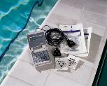
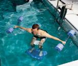
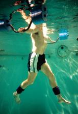
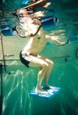
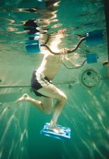
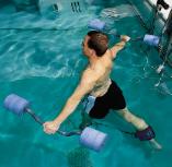
Brian Awbrey, MD and I recently published a research article showing the technique of aquatic biofeedback was valid and reproducible. It also showed the therapist could assess their patient’s progress quantitatively, allow the patient to effectively train while in the pool and ultimately, customize the treatment program to fit the individual needs of the patient.5 (Figure 1) Aquatic biofeedback utilizes the same techniques and procedures as its land-based cousin. Skin preparation and electrode placement are still important, however the critical step is the application of a waterproof barrier to seal the electrode site. (Figure 2) The waterproof barrier is the key to aquatic biofeedback. It enables the therapist the opportunity to specifically direct treatments and collect quantitative data while the patient exercises in the water. Since the surface EMG unit is hand-held and powered by a 9-volt battery, the threat of electrocution is eliminated and site infiltration by water remains nothing more than a momentary light tingle. The possibility of saturating an expensive surface electrode however remains a more realistic hazard. The patient performs the exercise routine while, ‘wired-up.’ The therapist directs and records specific events of the treatment session. Threshold-based uptraining of either unilateral VMO/VL or bilateral VMO’s is performed on certain exercises with emphasis placed on the quality of contractions, increasing magnitude of contractions, improving timing of recruitment and resolution of dysfunction and pain. The addition of aquatic therapy in the treatment of PFPS can provide an environment in which treatment can be initiated earlier, land-based exercises can be mastered sooner and specific skills can be advanced quicker. Aquatic biofeedback brings to the treatment regimen specificity, control, quantitative measurements and patient education. Athlete and armchair
artist alike will most likely suffer from PFPS at sometime in their lives.
Quick treatment and rapid resolution of the symptoms is imperative.
Aquatic therapy offers you the opportunity to treat them immediately;
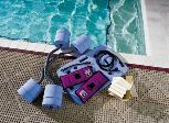
aquatic biofeedback allows you to treat them correctly. Exercises Phase I: 1-2 Weeks Goals: Increase quadriceps recruitment, restore normal patterns of muscle synergy, increase magnitude, improve timing of VMO recruitment vs. VL, increase ROM and decrease pain. Frequency: Two pool sessions and one land session per-week. Equipment: Thigh cubes, balance board, thigh/ankle cuffs with long and short cords (Figure 3), MyoTrac 2, sensors, bioclusive dressing (Figure 4). Note: Do not train through pain. Perform aquatic biofeedback on VMO and VL (or bilateral VMO’s). May require patella taping during pool session. Perform stretching exercises to appropriate muscles prior to exercises. Depending on the severity of symptoms, start with buoyancy assist and progress to buoyancy resist.
Exercises Phase II: transition Goals: Minimal to no pain with exercises, VMO/VL ratio 1:1 or better and full AROM. Frequency:
One pool session and two-land sessions per-week. Note: Progress from
small to large movements, slow to fast speeds and light to heavy resistance.
As skill develops, incorporate multi-joint control, add distractions,
randomize training activities and remove audio/visual feedback. Equipment:
Same as phase I
The featured exercise routine is initiated as soon as possible in conjunction with the land-based program. Progression from phase I to phase II is contingent on VMO output, balance between VMO/VL and pain scale with activity. Exercises may be added, modified or deleted as needed (or tolerated) in order to fit the needs of your patient, pool or time constraints.
Article reprinted with permission of the Sports Medicine Update magazine. |
|
|
|
|||
|
|||
|
|||
|
|||
|
|||
|
|||
|
|||
|
|||
|
|||
|
|||
|
|||
|
|||
|