
Recovery of the Post Operative Knee
The use of Electromyographic Biofeedback for Post Operative Quadriceps Femoris Muscle Recovery.
Vanessa Draper, Ph.d.
Introduction
Following an anterior cruciate ligament (ACL) reconstruction, immobilization and/or disuse of the operative limb can result in significant quadriceps femoris (QF) muscle atrophy and weakness, even within the first few days following surgery and certainly the first few weeks1. The focus of ACL rehabilitation programs is, therefore, the recovery QF muscle force and function (figure 1). Rehabilitative exercises during the early phases of treatment typically include QF muscle setting (QS) and straight leg raises (SLR). Often, these exercises are difficult to perform during the initial post operative weeks because of pain, edema, and possibly a disruption in normal joint receptor activity 2,3. If joint receptor feedback is distorted, the facilitory and inhibitory influences of this feedback on joint musculature are distorted and normal muscle contraction patterns become irregular and less effective. This may impede the performance of rehabilitative exersise and the recovery of muscle control and strength.
Several authors have suggested that EMG biofeedback(BFB) may be a valuable augmentor of receptor feedback from the knee musculature during QF exercise4,5,6,7. Via surface electrodes, motor unit activity from the QF in monitored during exercise and converted to a visual or audible signal.
While the patient may not be able to "feel" or perceive the muscle activity, he or she can see or hear the results of efforts to contract the muscle. Biofeedback has been shown to facilitate significant clinical improvements in cases of post operative hand rehabilitation8, shoulder joint instability9, spinal injury and stroke rehabilitation10,11 post meniscal repairs (12) and QF recovery following ACL reconstruction13,14.
EMG Biofeedback and QF Recovery
The recovery of QF muscle force following an ACL reconstruction first requires that the patient recover voluntary control of the muscle so that strengthening exercises can be performed effectively. The BFB is used to reeducate the patient in voluntary contractions of the QF, providing the patient with immediate information regarding the correctness of their efforts during QS's and SLR's. This information not only augments distorted or diminished feedback from the joint receptors but, in addition, it provides a motivational element. Performance assessment has been described as an important source of motivation. When individuals are more keenly aware of their performance level, they are more impelled to set and strive for goals. When patients are provided with a visual or auditory representation of otherwise covert or masked muscle activity and are given a qualitative goal, exercise effort and outcome are enhanced.
Treatment Program
Patients may begin working with biofeedback as soon as possible after surgery. Typically, patients are asked to perform QS's and SLR's within a day of surgery and they can easily begin the BFB monitoring at this time.
Using the MyoTracTM EMG System
The MyoTracTM single channel EMG system from Thought Technology Ltd. is a source of visual and auditory feedback that allows the patient to monitor the precision and intensity of each muscle contraction. Variable settings on the unit allow the therapist to monitor progress as well as set future goals for the patient. In order to develop an optimal rehabilitation plan, it is necessary to determine the level of activation that the post operative quadriceps muscle is capable of producing following surgery. Prior to the first session reading, several adjustments must be made to the unit as follows:
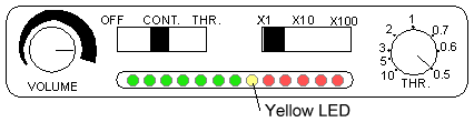
Figure 1. External Panel Settings
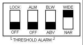
Figure 2. Internal Panel Settings
- Choose X1 for the scale reading to utilize the primary level of 0-20 uV.
- Set the threshold switch to continuous (CONT) in order to allow the patient to see/hear any changes in the muscle activity during the exercises.
- Set threshold to above (ABV) to provide only visual biofeedback until the threshold level (yellow LED) has been exceeded.
- Set the threshold dial to the lowest setting of 0.5. Adjust the volume level.
- Plug the sensor cable into the appropriate jack and secure the electrodes on the subject as follows:
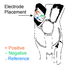
Figure 3. Electrode Placement
With the leg in extension place the electrode 3-5 cm above the superior border of the patella and 2-3 cm medially (figure 3), making sure to have the positive and negative electrodes parallel to the muscle fibers. The incision pattern will determine the actual location but the goal is to focus on extensor mechanism activity during QS's and SLR's. The sensor head should be taped or fastened to the site. Many therapists find that self-adhesive triodes give optimal readings, however, care must be taken to avoid further irritation to any already sensitive areas.
Technique for EMG Training
The patient is first trained to use the BFB with the QS exercise. With the patient sitting up, affected leg straight out on the table and electrodes in place, check the LED's on the MyoTracTM while the patient is relaxing the QF. If, in this position, the red LEDs are lighting up, adjust the threshold dial counter-clockwise until the green lights are activated. Should the highest threshold setting fail to move from the red LED area to the green LED's, switch the internal settings to X10, return the threshold dial to 0.5 and recheck the LED's. Repeat this procedure using the X100 if necessary. The more advanced the muscular atrophy, the lower the required threshold settings. Once the appropriate settings have been established, the patient may begin strengthening exercises for the QF.
Beginning with the LED in the green, the patient should start QS while watching the LED and/or listening to the auditory signal from the MyoTracTM. Maximal effort should be required for the red lights to be activated. By readjusting the settings, the difficulty of an exercise may be increased or decreased, according to the needs of the patient. It is important to stress, however, that the importance lies in the quality of contraction versus the quantity.
Should the patient have difficulty initiating a contraction, several verbal cues may help, including "press the back of your knee into the table" or "look for the knee cap to shift and the foot to raise". Once enough activity is generated to register and be displayed on the BFB unit, this feedback signal can be used to guide further efforts to contract (MyoTrac will detect signals from .08 to 2000 uV and, in our experience, most postoperative patients will initially register approximately 10-20 uV). The patient should be able to perform 3 sets of 10 QS's to the satisfaction of the therapist before moving on to the next phase of the program in order to avoid improper technique. In order for the BFB to be used as a training tool, low goal levels of activity should be set and met before setting higher goals.
The mistake that most patients make during initial efforts to contract the QF and straighten the leg is co-contracting the hamstring (HS) muscles in a compensatory manner. While the patient may feel that they are exerting enough effort to straighten the limb, the minimal activity recorded from the QF and displayed on the BFB unit will make them aware that their efforts are made with the wrong muscle group. We recognize that many therapists teach co-contracting as a means of strengthening the knees following surgery, however, even with co-contracting, the QF must contract effectively for knee extension to take place. Biofeedback can easily be adapted to this exercise. It would also be advisable to use a dual channel unit or 2 single channel units in this case and monitor both HS and QF activity during knee extension.
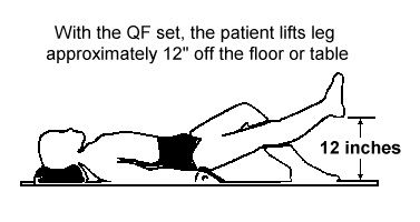
Figure 4. Straight Leg Raise Exercises
Once BFB can be used successfully with the QS exercise, the patient then uses it with the SLR's. This exercise is performed in a supine position, leg extended. The patient sets the QF first and then lifts the leg approximately 12 inches off the floor or table and holds for a 5 second count (figure 4). By monitoring the LED's during each SLR, the patient will be able to assess the quality of the QF contraction. The QF should remain set with no evidence of extensor lag during the hold. Monitoring this exercise with the BFB makes the patient more aware of changes in the QF motor unit activity during the hold time and better able to maintain a constant and complete contraction throughout the exercise repetition.
Conclusion
In our experience, BFB is most useful during the first 2-4 weeks following ACL reconstruction. We suggest that it be used with QS's (3 sets of 10 repetitions, 3 times per day) and SLR'S (3 sets of 10 repetitions 3 times per day; progressively adding 2-10 lbs of ankle weight, as tolerated). Thresholds should be set so that the patient has to exert maximally to achieve the goal. Patients should be encouraged to increase these thresholds as soon and as regularly as possible, otherwise the BFB becomes only a monitoring device and not a training tool. It is important to remind the patient that actual microvolt values may vary according to the degree of edema present around the recording site as well as to variations in electrode placement. Patients may be falsely encouraged or discouraged if they rely too much on actual values. The values will, however, be relative to the accuracy and intensity of the exercise effort and this information can be extremely helpful, as well as motivational, for the patient rehabilitating the post operative knee. The more feedback that the patient receives about the quality of their muscle contraction, the more control they will have over the retraining of that muscle, and in turn, will be more motivated to continue therapy. BFB is proving to be a key in providing the therapist with important information on the patient's progress, as well as helping the patient monitor his/her own success.
References
- Noyes FR, Mangine RE, Barber S: Early knee motion after open and arthroscopic anterior cruciate ligament reconstruction. Am J Sports Med 15:149-159, 1987.
- deAndrade JR, Grant C, Dixon A St. J: Joint distension and reflex muscle inhibition in the knees. J Bone Joint Surg (Am) 47:313-322, 1965.
- Krebs DE, Staples W, Cuttita D, Zickle R: Knee joint angle, It's relationship to quadriceps femoris muscle activity in normal and post-arthrotomy limbs. Arch Phys Med Rehabil, 64:441-447,1983.
- Soderberg GL, Minor SD, Arnold K et al: Electromyographic analysis of knee exercises in healthy subjects and in patients with knee pathologies. Phys Ther 67: 1691-1696, 1987
- Hald RD, Bottjen EJ: Effect of visual feedback on maximul and submaximal isokinetic test measurements of normal quadriceps and hamstrings. Journal of Orthopedic and Sports Physical Therapy 9:89-93, 1987
- Delitto A, Rose SJ, McKowen JM, Lehman, RC, Thomas JAS, Shively RA: Electrical Stimulation versus voluntary exersise in strengthening thigh musculature after anterior curciate ligament surgery. Phys Ther 68:660-663, 1988.
- Lucca JA, Recchiutu SJ: Effect of electromyograhic biofeedback on an isometric strengthening program. Phys Ther 63:200-203, 1983.
- Brown DM, Nahai F: Biofeedback strategies of the occupational therapist in total hand rehabilitation. In Basmajian JV(ed): Biofeedback: Principles and Practice for Clinicians. Baltimore, MD, Williams and Wilkins, 1983, pp 90-106.
- Beall MS, Diefenbach G, Allen A: Electormyographic biofeedback in the treatment of volunteer posterior instability of the shoulder. Am J Sports Med 15: 175-178, 1987.
- Nacht MB, Wolf SL, Coogler CE: Use of electromyograhic biofeedback during the acute phase of spinal cord injury. Phys Ther 62: 290-294, 1982.
- Wolf SL: Electromyographic applications to stroke patients: A critical review. Phys Ther 63: 1448-1459, 1983.
- Sprenger C, Carlson K, Wessman H: Applications of electromyographic biofeedback following medial menisectomy. Phys Ther 59: 167-169, 1979.
- Draper V: Electromyographic biofeedback and the recovery of quadriceps femoris muscle function following anterior cruciate reconstruction. Phys Ther 70: 11-17, 1990.
- Draper V, Ballard L: Electrical Stimulation versus electromyographic feedback in the recovery of quadriceps femoris muscle function following anterior cruciate ligament surgery. Phys Ther 71: 455-464, 1991.
Copyright, 1997 The Biofeedback Federation of Europe
Ā