
The Unstable Shoulder
Biofeedback Training of the External Rotators to Centralize the Humeral Head in Patients with Anterior Shoulder Instability and/or Pain.
Linda Saboe, B.P.T., M.C.P.A.
Judy Chepeha, Bsc.P.T., M.C.P.A.
David Reid, M.D., M.Ch.H., F.R.C.S.
Gary Okamura, M.D.
Michael Grace, Ph.D, P. Eng
The Glen Sather Sports Medicine Clinic, and the Division of Orthopaedics.
The University of Alberta
Introduction
Anterior shoulder instability and impingement are common athletic complaints associated with overuse, joint laxity, post-traumatic dislocation and muscle imbalance. While traditionally treated as clinically discrete entities, it is now accepted that considerable overlap exists between functional instability and anterior impingement(1-3).
Until recently, rehabilitation programs have emphasized subscapularis strengthening on the assumption that this muscle provided an anterior buttress, preventing anterior humeral head subluxation(4-6). Turkel (1981) has demonstrated inability of the subscapularis to cover the anterior humeral head in abduction and external rotation(7) and Garth reports that internal rotation forces actually contribute to anterior displacement(2). These findings provide an explanation for high failure rates of traditional rehabilitation programs(8-11).
Jobe and Perry's electromyograhic work identifies the external rotators, and in particular, the infraspinatus, to be the primary dynamic anterior shoulder stabilizers in abduction and overhead motions(12-14). This dynamic stability is provided by preventing forward motion of the humeral head in the glenoid fossa.
In 1988, a treatment protocol utilizing single channel electromyographic biofeedback was developed; it has been continuously tested and enhanced through controlled clinical trials at the University of Alberta. This program utilizes targeted muscle feedback to perfect motor skills. By electronically monitoring and amplifying activity of the external rotators during an apprehensive motion with immediate visual and auditory feedback to the subject, the performance is changed or shaped. This program, which emphasizes muscle control rather than strength, requires motivation, training, and lifelong routines to maintain the established engram and control shoulder stability.
USING A SINGLE CHANNEL EMG BIOFEEDBACK SYSTEM
The MyoTrac single channel EMG biofeedback unit, from Thought Technology, is valuable in the reinforcement of appropriate external rotator activity. Patients are provided with visual and auditory feedback of appropriate muscle activity. The unique MyoScan sensor amplifies the muscle signals at the pickup site, thereby providing excellent sensitivity with no electrical interference (see figures 1 and 2)
SINGLE CHANNEL BIOFEEDBACK TREATMENT PROGRAM
- Electrode placement is critical. Using the disposable triode electrodes, attach the sensor below the scapular spine with the 2 active electrodes parallel to the orientation of the muscle fibers. Do not place it over the posterior deltoid as increased activity in this muscle would drive the humeral head anteriorly. The patient remains connected to the biofeedback unit during training and must practice at home, both with and without the unit. For home practice, the therapist might wish to place an indelible mark on the skin for electrode placement.
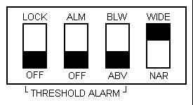
Figure 1. Inside Pannel settings
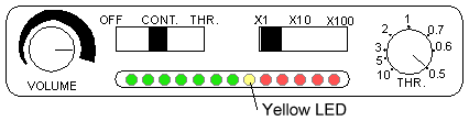
Figure 2. Threshold and Gain settings
- To determine threshold and gain settings, have the patient flex the shoulder forward to 70 degrees with the gain switch at X1, and turn the threshold control until the yellow LED illuminates. If the activity is greater than 10uV at 70 degrees, set the gain settings to X10. Again, have the patient flex the shoulder forward to 70 degrees while turning the threshold control until the yellow LED illuminates.
- Ensure the shoulder is in a pain-free neutral position, the threshold switch is set to 'CONT', the volume is set at a pleasant level (with or without the earphones) and the threshold control and gain reading switch remain in the positions set previously in step 2. Instruct the patient to use the visual and audio feedback to increase EMG activity well above the yellow LED. This is done by tightening the rotator cuff muscles in the neutral position in order to glide and hold the humeral head posteriorly (figure 3). This is a key component and must be successfully performed 100 times (ten sets of 10) prior to progressing to active movement. The use of many repetitions builds endurance. This procedure is quite fatiguing; it may require several sessions before the patient can progress to step 4.
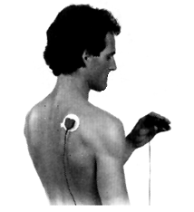
Figure 3. Teach contraction in neutral position
- With the theshold set at twice the value achieved in Step 2, place the patient's elbow in flexion (figure 4). Instruct to forward flex the adducted and neutral rotated shoulder to 90-100 degrees. As the shoulder is flexed between 70 and 90 degrees, have the patient tighten the rotator cuff and achieve the threshold setting, trying to light-up the lights to the right of the first yellow one. If pain or a sense of subluxation is experienced, stop, rest and start again through a smaller arc of movement and/or with reduced threshold settings. When the patient can successfully perform 100 consecutive repetitions, progress by increasing the threshold and/or movement as shown in figure 5.
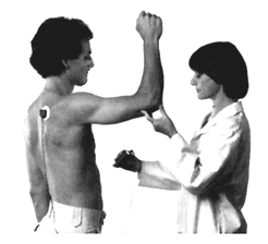
Figure 4. Forward flex to 90-100 degrees
MOVEMENT PROGRESSION
As the patient masters each level, progress through the following exercises:
a) Forward flexion with a straight elbow.
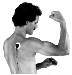
Figure 5.
b) Forward flexion with increasing external rotation.
c) Abduction with flexion progressing to elbow extension
d) Abduction with elbow extension with increasing external rotation.
e) Abduction from flexion.
f) Abduction from flexion with increasing external rotation.
g) Reach for objects behind back or overhead.
When the above progression of increasingly difficult tasks has been completed, progress to the activities specific to the sport or work task that caused the difficulty. Break the movement down into component parts and introduce catching or throwing activities in preparation for a gradual return to normal activity (figures 6, 7).
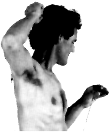
Fig. 6.
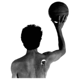
Fig. 7.
OTHER EXERCISES
If general weakness exists, instruct the patient in appropriate progressive resistance exercises. Include pushups for serratus anterior (with the arms abducted) and external rotation exercises resisted using surgical tubing. Avoid resisted exercises which load in an impingement position (figure 5). All pain free activities are allowed and encouraged. The patient will require two to three weeks of supervised physiotherapy, but must do a life-long home program to maintain the engram. It might be desirable for patients to return for occasional brief refresher courses.
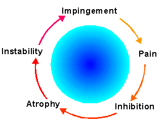
Figure 8. Instability and impingement are related
CONCLUSION
This program emphasizes muscle control. Strength acquisition is important, but secondary. Electrode placement is critical. The biofeedback program is physically and mentally demanding, therefore, appropriate rest periods and encouragement must be provided. Slow and careful progression is usually necessary. Commitment by the therapist and client are required for the program's success.
REFERENCES
- Rowe CR: Factors related to recurrences of anterior dislocation of the shoulder. Clin Orthrop 20:21, 1961
- Garth W, Allman F. Armstrong W: Occult anterior subluxations of the shoulder in noncontact sports. Am J Sports Med 15:579-585, 1987.
- Reid D, Saboe L, Burnham R: Current research of selected shoulder problems. In: Donatelli R. (Ed.) Physical Therapy of the Shoulder. Churchill Livingston, New York, 1987
- Magnusson PB: Treatment of recurrent dislocation of the shoulder. Surg Clin N Am 25:14-20, 1945.5 Adams JC: Recurrent dislocation of the shoulders JBJS 30B(1) 26-38, 1948.
- De Palma AF: Factors influencing the choice of a modified Magnusson procedure for recurrent anterior dislocation of the shoulder. Surg Clin N Am 43: 1647-1649, 1963.
- Turkel S, Ithaca M, Panio M, et al: Stabilizing mechanisms preventing anterior dislocation of the glenohumeral joint. JBJS 63A:1208-1217, 1981.
- Rowe C. Zarins B: Recurrent transient subluxation of the shoulder. JBJS (Am) 63A:863-871, 1981.
- Hastings D, Coughlin L: Recurrent subluxation of the glenohumeral joint. Am J Sports Med 9:352-355, 1981.
- Simonet W, Cofield R: Prognosis in anterior shoulder dislocation. Am J Sports Med 12:19-24, 1984.
- McLaughlin HL, Cavallaro WU: Primary dislocation of the shoulder. AM J Surg 80:615-621,1950.
- Perry J, Anatomy and biomechanics of the shoulder in throwing, swimming, gymnastics and tennis. Clin Sport Med 2(2):247-270, 1983.
- Jobe F, Tibone J, Perry J, et al: An EMG analysis of the shoulder in throwing and pitching. Am J Sport Med 11:3-5, 1983.
- Gowen I, Jobe F, Tibone J, et al: A comparative electromyographic analysis of the shoulder during pitching. Am J Sport Med 15:586-590, 1987.
Copyright, 1997 The Biofeedback Federation of Europe
Ā